Deep Image Segmentation of Medical Data
Cardiac MRI derived biventricular mass and function parameters, such as end-systolic volume (ESV), end-diastolic volume (EDV), ejection fraction (EF), stroke volume (SV), and ventricular mass (VM) are clinically well established. Image segmentation of the human heart can be challenging and time-consuming, due to its complex anatomy. Given an MRI measurement of the upper body, the objective is to automatically delineate the endocardium and epicardium of the left and right ventricle for subsequent volumetry.
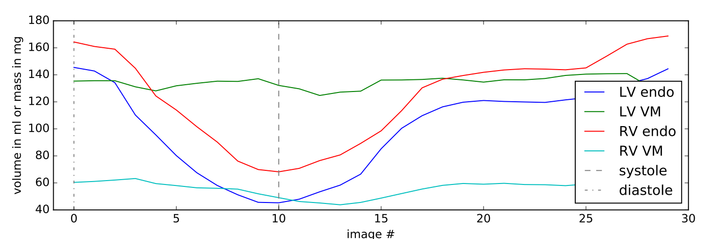
Segmentation based volumetry allows for fast and high-precision estimation of clinical parameters over a whole cardiac cycle. Time-resolved determination of cardiac volumes enables the development of novel approaches for the analysis of cardiac diseases and has manifold applications in preventive medicine.
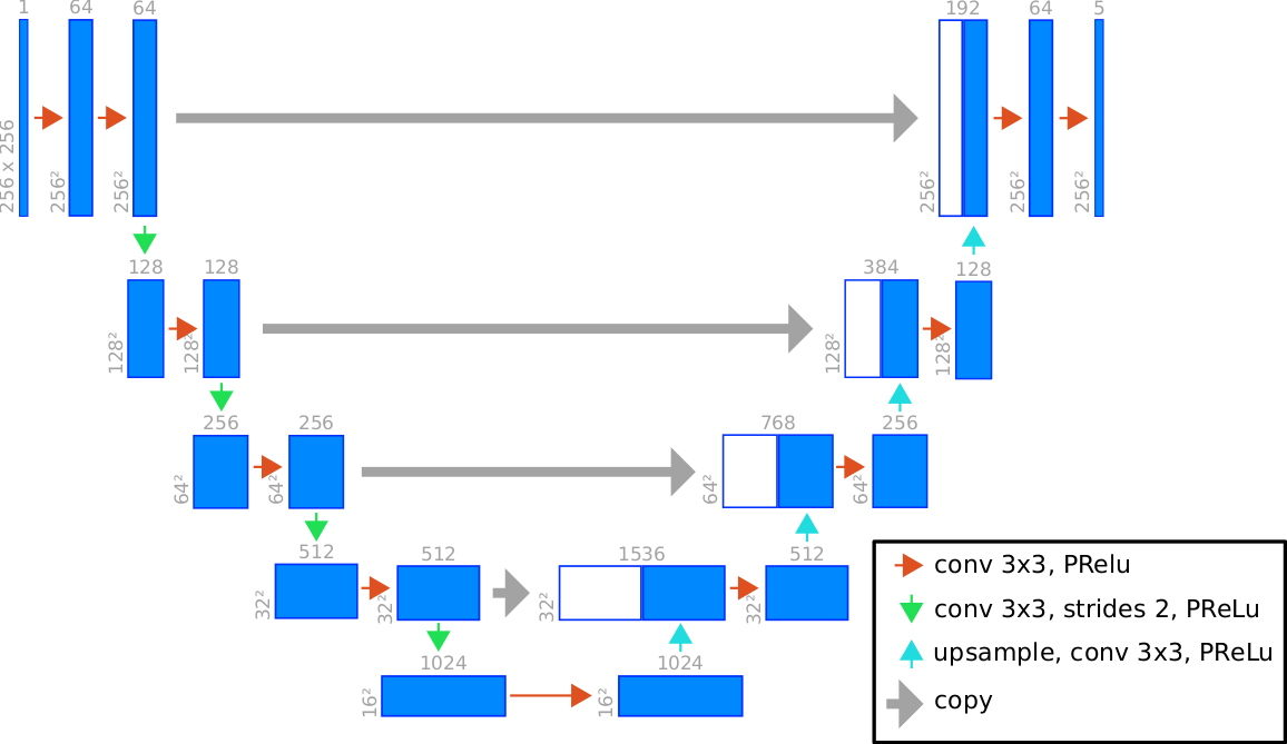
We introduce \(\nu\)-net ("new net"): a state-of-the-art deep segmentation approach facilitating automated segmentation of the left and right ventricle which is an extension of the traditional U-net architecture. \(\nu\)-net performs multi-scale pattern detection with the help of hierarchical nested convolutional layers. Hence, cardiac features can be reliably mapped onto binary segmentation masks in low contrast MR images.
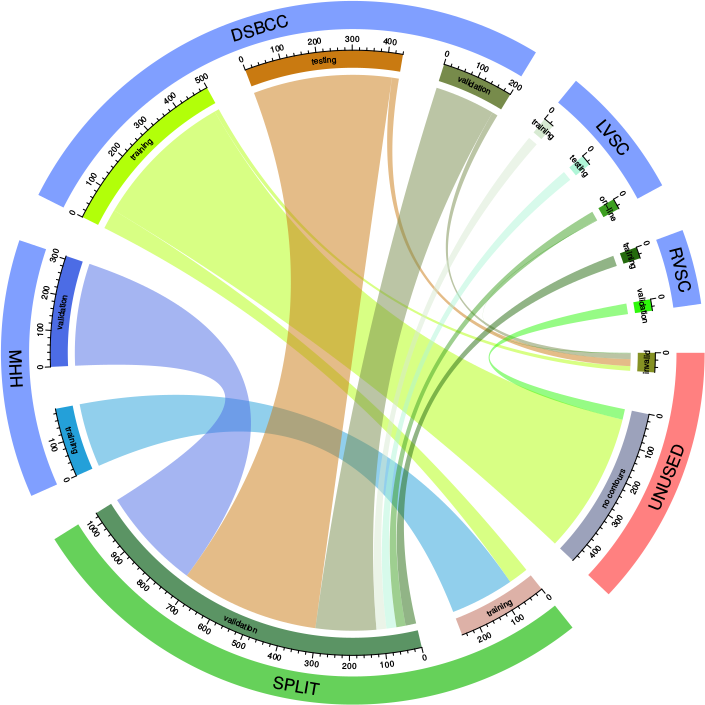
The \(\nu\)-net framework exhibits excellent generalization properties on new data produced by a wide range of MR scanners since it was trained on an extensive collection of MR images stemming from independent data sets. This allows for a straightforward application with no or few fine-tuning steps. This robustness is achieved by means of a comprehensive training protocol covering four distinct data sources consisting of hundreds of cardiac scans:
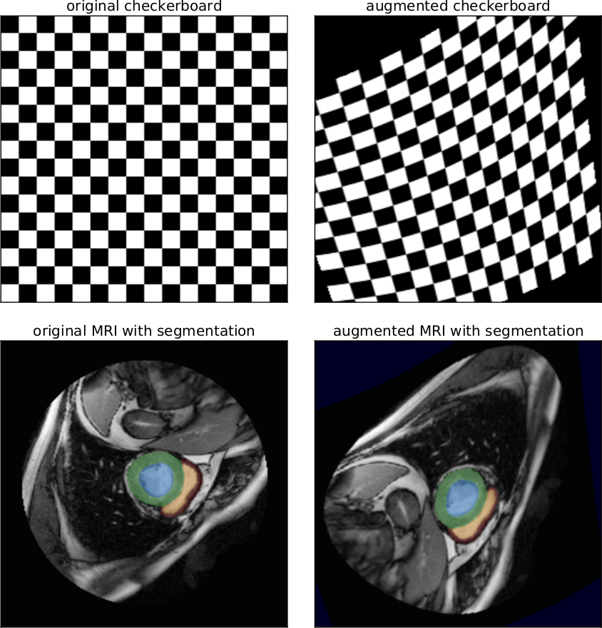
Further robustness is achieved by employing non-linear deformations to the spatial domain of MR images. Hence, the \(\nu\)-net architecture is capable to robustly detect cardiac features independent of their spatial orientation, potential distortions and variations introduced by the acquisition technique of a certain MR scanner.